
Endodontic treated teeth are avital, even so their survival in oral medium must be as well as vital teeth. Therefore the need of assessment and analyzing the endodontic procedure and methods of canal obturation is very important esthetic and functional segment of our work.
The aim of this research project is to analyze and evaluate canal obturation in correlation with different techniques of conditioning the dentine in the root canal and different techniques of gutta-percha application.
Different segments will be an object of analysis: These specific goals will be analyzed
to analyze canal obturation and adhesion of sealer depending from conditioning of dentin walls (Nd:Yag laser and 17% liquid of EDTA)
to evaluate the adhesion of sealer on apical, middle and coronal third of the root canal
to evaluate the quality of canal obturation with GuttaFlow gutta-percha technique
to evaluate the quality of obturation when using modified conventional gutta-percha technique
to analyze and compare the established parameters and to notice their role in successful tridimensional canal obturation
MATERIAL AND METHOD
This project will be realized on 120 single - rooted human teeth, extracted from orthodontic or prosthetic reasons. According to technique of gutta-percha application teeth will be divided in two groups of 60 teeth in each:
- First group – 60 endodontic treated teeth obturated with AH plus sealer and Gutta Flow technique
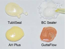
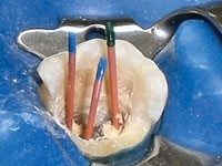
- Second group – 60 endodontic treated teeth obturated with AH plus sealer and modified single cone gutta-percha technique
In each group will be performed cleaning of smear layer before definitive canal obturation divided in two groups:
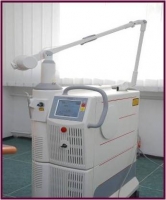
- Subgroup A – 30 teeth where root-canal dentine will be conditioned with Nd:Yag laser – emission in three series of 5 seconds
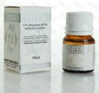
- Subgroup B – 30 teeth in which smear layer will be removed with applying 17% EDTA for 3 minutes
-
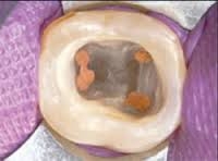
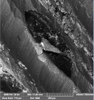
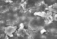
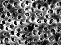
Quantitative and qualitative analysis and evaluation of canal obturation and its marginal adaptation will be performed on Scanning Electron Microscope by two irrespective teams of researchers involved in survey.
Applicative implementation
Obtained data from the experimental part of this project will be used for making a doctrine standpoint and protocol for processing of root canal obturation. Implementation will be valorized in restorative dentistry, endodontics, oral surgery, pedodontics and prosthodontics. Improving the success of endodontic treatment as well affects on functional and esthetic restoration of teeth.
Проф. д-р Ивона Ковачевска – главен истражувач
Проф. д-р Цена Димова – истражувач
Проф. д-р Златко Георгиев – истражувач
Доц. д-р Илијана Муратовска – истражувач
Доц. д-р Киро Папакоча – млад истражувач
Д-р Ефка Жабокова Билбиловска PhD – млад истражувач
Ас. д-р Ана Радеска – млад истражувач
Ас. д-р Наташа Денкова – млад истражувач
Ас. д-р Сања Нашкова – млад истражувач
Д-р Михајло Петровски – млад истражувач
Д-р Катерина Фотева – млад истражувач